When Less is More, Regarding Radiation

J-Martin-Palomo-Headshotby Juan Martin Palomo DDS, MSD

We all know Cone Beam Computed Tomography (CBCT) by now. It allows a non-invasive, usually less than 10-second capture of the craniofacial anatomy, which is able to create all possible traditional 2D radiographs, giving far more diagnostic information. Probably the main mentioned reason of why it has not replaced the traditional panoramic and cephalometric radiographs in clinical orthodontics, is radiation. Depending on the settings used, it could expose the patient to more radiation than that of a ceph and pano. The radiation would still be considered low, according to the American College of Radiologists, and is less than the additional annual cosmic radiation that somebody living in high altitudes, such as Colorado, receives, but nevertheless, more than a ceph and pano. But this is no longer the case, for a while.

Most major CBCT manufacturers have now a low-dose scanner in the market, which allows the 3D image to be captured, with less radiation than a panoramic radiograph. It givless is more Palomo
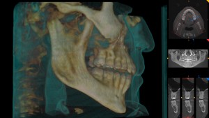
es a complete 3D image, where a pano, ceph, and complete 3D view can be used, for less radiation than that of a distorted pano. How is this possible? The answer is “capturing technology”.

When a low dose 3D image is captured, the scanner does not go all 360 degrees around the patient’s head. Sometimes it is just 180 degrees. The 3D image is basically a combination of several static images (radiographs) taken while the scanner rotates around the patient’s head. In a low dose option, the number of images captured are less than 200, as opposed to the usual 300-600. When a panoramic radiograph is being taken, the x-ray beam is “on” the whole time, beeping, while going around the patient’s head. In a low dose CBCT scanning, pulse technology is used, so the x-ray beam is only “on” for a fraction of a second while taking a quick image, turning itself “on” and “off” automatically, resulting in a total radiation exposure of about 2 seconds.

All this in combination with low radiation settings, results in total effective radiation to the patient in the teens or low twenties, compared to high twenties for a pano. And in this numbers game that is effective radiation, the suggested safety threshold is 30 microsieverts. Anything below 30 microsieverts can be considered low, and fair game.

Now for the very necessary disclaimers. This does not mean that because we can get to less than 30 microsieverts we should scan everybody. Zero is still less than 30. So if a radiograph that can answer the question clinically posed has already been taken, retrieve it and do not take an additional one. If no radiograph is necessary, don’t take one just because you can. But if a radiograph is needed, and a low dose CBCT is an option, I would have a hard time justifying a ceph and pano, as opposed to a low dose CBCT. And to take a pano or ceph before taking a low dose CBCT would also be unnecessary additional radiation to the patient.

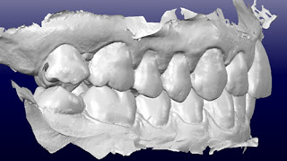
The low dose CBCT alone has more than sufficient image quality for what we need in orthodontics. I have seen images from several manufacturers, and this is clearly a situation where less radiation to the patient is also more information to the doctor. What better win-win that this can we ask?

Nanotechnology: From Small Scale to Great Innovations

By Dr. Celestino Nobrega

dr Celestino Nobrega 2007Are you prepared for the amazing benefits and innovations that Nanotechnology will shortly bring to orthodontics? Richard Feynman (http://www.feynman.com), an American theoretical visionary physicist, introduced Nanotechnology as a science that embraces the capability to see and to arrange atoms and molecules according to a particular convenience or goal.

Nanotechnology involves the development and utilization of structures, devices, and systems that have properties and new functions due to their small size. A nanometer is one-billionth of a meter. For reference, consider that there are about 25,400,000 nanometers in an inch, and a single sheet of paper is about 100,000 nanometers thick.

The NNI (National Nanotechnology Initiative) is a U.S. Government research and development initiative that is focused on delivering the shared vision of “a future in which the ability to understand and control matter at the nanoscale leads to a revolution in technology and industry that benefits society.” NNI is chartered to develop a framework for sharing strategies in order to support nanoscale projects. Common goals, strategies and priorities are now being drawn for distinct science fields, such as biology, engineering, chemistry and, of course, materials science. With this support, nanotechnology R&D is taking place in academic, government, and industry laboratories across the United States.

As Orthodontics progresses into a refined science and with the support of technology advancements, unimaginable results can be achieved in the near future, especially when our specialty can leverage nanotechnology innovations such as selective biosensors. The oral cavity can be considered as an important source of information that could be extremely helpful not only for orthodontic treatment, but also for early stage diagnosis and monitoring of systemic diseases. It’s largely known that the exhaled human breath contains several Volatile Organic Compounds biomarkers (VOCs). Accurate detection of these VOCs can provide essential information for the diagnosis of those diseases. For example, Acetone (CH3COCH3), H2S, NH3, NO, and Toluene can potentially be used to evaluate diabetes, halitosis, kidney malfunction, asthma, and lung cancer, respectively.

Breath analysis, pH level and temperature data can be captured and processed by multiple sensors and could potentially reduce the medical diagnostic costs for patients suffering from chronic illnesses. In addition, patients’ quality of life could be improved. For example, diabetic individuals could possibly benefit from using non-invasive nanostructured hemitubes Silicon-doped (WO3) films to sense acetone exhaled breath levels, which can eliminate painful and invasive fingertip pricking.

As I observe the emergence of recent technology advancements within orthodontics, I can envision a future of innovative orthodontic portable devices that can accurately capture, track and transmit these previously mentioned biological signals.

In this category, we can include the exciting new innovation of dental movement acceleration devices (periodontal tissue activation by vibration). Along with achieving their primary objective of delivering pulsatile forces to accelerate tooth movement and to reduce treatment time, these devices could also be used to capture intra-oral data through the action of nanosensors and wirelessly transmit this information to personal mobile devices and laptops. The remotely collected data can be stored on the cloud to create an intelligent system for support of clinical decisions. This robust wireless communication and database creation has the potential to support multiple users throughout the orthodontic treatment process:

  • Orthodontist: electronic health records (EHR) enrichment; treatment plan refinement according to patient features; suggestions for treatment plan improvement according to case’s evolution; warning signals and alerts to monitor patient’s general and intra oral health.
  • Patient: real time communication with the Orthodontist or their staff through smartphone connection; treatment status check; sharing treatment experiences with other potential patients; automatically get FAQ answers.
  • Companies, industries and laboratories: helpful database for new products designs, services and needs.
  • Scientific research: Database for systematic reviews, Meta-Analysis.

So, are you prepared for “small” technology and big changes?


3D Printing and Orthodontics

By Dr. Christian Groth

As we move towards the 2015 AAO Annual Session in San Francisco many of us will be making check lists for items to investigate at the exhibition hall and lectures. Anybody who has attended recent meetings has seen that intraoral scanners are a hot topic. Every year new products are being released, or updates to current systems are offered. Intraoral scanning has opened the door for additional technologies within (and outside of) the orthodontic office. Dr. John White wrote a very informative blog post in February talking about the use of intraoral scanning for same day consultations (click here to read it). As more people are offering clear aligner therapy (including general dentists and the mail order aligner system that we all know about) it is time that we differentiate ourselves as orthodontic specialists. One way in which we can do this is to incorporate 3D printing into our daily practices.

3D printing, also known as additive manufacturing, is a process by which a physical object is created from a digital file (check out a video of 3D printed models here). There are several different types of 3D printers available that range in price from a few hundred dollars to almost one hundred thousand dollars. They all have one thing in common: they build models layer-by-layer with a build platform that moves vertically. The smaller the layer thickness the better looking the model will be. The four most popular types of printers are: Fused Deposition Modeling (FDM), Stereolithography (SLA), Digital Light Projector (DLP), and Polyjet Photopolymer (PP). Without getting too technical here is how each basically works. FDM involved heating up a thin strand of plastic resin that comes off of a spool and is deposited in layers as thin as 100 microns. SLA and DLP technologies are similar in that they utilize a vat of liquid, photosensitive resin. When the light hits the resin it is cured and platform moves to enable the next layer to be cured. The different between SLA and DLP is that SLA uses a single laser point to draw an image whereas DLP uses a projected image to cure a whole layer simultaneously, which allows the printing process to move faster (think of this as the difference between drawing a picture and stamping a picture). PP printers are probably the most popular in dentistry and use inkjet technology (yes, just like your desktop printer). Liquid resin is jetted out of nozzles in an extremely accurate fashion and cured by a UV. Layer thickness of SLA, DLP, and PP printers can be as low as 16 microns (for your reference the average piece of paper is 100 microns thick).

While the technologies differ between printers what truly sets them apart is the quality of the parts. Cheap printers are made from cheap parts that can degrade over time and result in inaccurate models. It is truly a case of you get what you pay for. So if you are in the market for a 3D printer to be used in your practice, buy the best one that you can afford.

3D printed models can be used for anything in the orthodontic office.   The most practical use is for retention and relapse treatment. A major downside to stone models is that they are often destroyed during the retainer fabrication process. 3D printed models do not get ruined and can be used as many times as necessary for retainer fabrication. Imagine being able to print a model, make a clear retainer, and mail it off to a patient who is away at college. What a service you have just provided to your patient and they never stepped foot in your office! Pairing 3D printing with one of several software programs available allows us to create sequential setups/models for minor tooth movement. By controlling the process we control the overhead and thus have the ability to pass these savings on to the patient. Another great service that we can offer our patients if they have a lapse in retainer wear.

Whatever your practice is like there is a place for 3D printed models in it. While you will pay a little bit more for the physical model, the longevity, versatility, patient excitement, and ability to virtually eliminate alginate impressions from your practice will pay off in the end!

The Digital Generations

By Anthony M. Puntillo DDS, MSD

Dr.-Puntillo-PictureThe majority of the U.S. Workforce today is comprised of three generations:  Boomers (1946-64), Xers (1965-80) and Millennials (1981-99), each generation with its own unique set of characteristics.  The American Association of Orthodontists (AAO) now reports that more than 51% of its membership is composed of Gen Xers and Millennials.  By virtue of their birth timing Xers and Millennials, including myself (1966), were the first generations to grow up with computers in their homes.  Although Gen Xers differ from Millennials in many ways, technology is now ingrained into nearly every part of both generations’ lives.  For those Xers and Millenials that also happen to be orthodontists, this attachment to technology includes not only their personal lives, but also their orthodontic practices.

Over the last few years, my blog posts have centered on the discussion of a “Digital Orthodontic Practice.”  A digital practice must include not only the management and record keeping aspects (paperless) of our offices, but also clinical diagnosis and tooth alignment functions.  In this post, I want to highlight the current opportunities for moving digital in the clinical portion of your practice.

Diagnosis:

The clinical care for most orthodontic patients begins with a diagnosis and a treatment plan.  Given that Kodak is now only a shell of the company that it once was, I think it is safe to say most orthodontic practices are now taking digital photographs, instead of film, as part of their diagnostic records.  The recent 2014 JCO study of Orthodontic Diagnosis and Treatment Procedures1 found that more than 91% of the respondents used digital radiography, 69% used CBCT either routinely or occasionally, 41% used digital models and 28% used intraoral digital scanners.  Additionally,  the American Board of Orthodontics (ABO) recently announced that all initial models for their exam must be submitted in a digital format.  While the JCO survey included a relatively low number of respondents (n=135), I believe the findings are indicative of the Electronic Health Record (EHR) movement in all of the health care profession.  This movement, aided by government mandates and subsidies, has now breached the threshold level.  The train has left the station.  If you and your practice intend to stay relevant over the next decade, you absolutely need to be utilizing digital diagnostic records.

Tooth Alignment:

As our profession transitions to a digital diagnostic record norm, some are looking to move beyond diagnosis to digitally construct tooth aligning appliances.  In 1999, Align Technology opened the door to digital orthodontic tooth alignment with the introduction of the Invisalign system.  The system at that time relied upon traditional dental impressions, but today intraoral scanners and 3D printing have allowed for the elimination of the impression procedure.  Whether it be Align, or any other current Clear Aligner option, a digital model (.STL) of a patient’s dentition can be captured with a scanner, the teeth can be aligned using computer software, and treatment appliances (clear aligners) can be fabricated by machines based off of the digital “plan”.  Furthermore, this process can now also be utilized for patients using traditional bonded brackets.  Custom brackets along with custom bracket placement jigs and custom wires digitally planned and robotically bent are possible.  In large part because of costs and the learning curve, the digitization of clinical orthodontic procedures has not yet been completely accepted.   However, as the techniques become more refined, we should expect the cost to include them into our practices to decrease and implementation by the tech savvy Xers and Millennials to accelerate.  If you are an Xer or a Millennial, and have not already incorporated digital tooth alignment into your practice, you should be planning to do so in the near future.  If you are a Boomer, and potentially less comfortable with technology, you need to consider if you can afford to ignore this change.

Creating an esthetically pleasing and stable smile, can be a bit like designing and constructing a building.  In a recent conversation with a Boomer architect friend of mine he described the digital changes his profession has undergone.  My friend reported that my office, built in the year 2000, was one of the last buildings he drew by hand.  All of his projects now are digitally designed using 3D CAD technology, allowing him to plan and visualize the end construction result more effectively.  The transition in the architectural profession took time and learning.  Change is never easy.  However, as my friend now approaches the end of his career, he finds the “old” way inefficient and less accurate.   Whatever generation you were been born into, I encourage you to embrace the digital change our profession is in the midst of.  I am certain a digital orthodontics will ultimately benefit you and your patients.

1Keim Et.Al. 2014 JCO Study of Orthodontic Diagnosis and Treatment Procedures, Part 1: Results and Trends Journal of Clinical Orthodontics 2014; 48:10 pages 607-630.

Virtual Setups Using Intra Oral Scanners for Same Day Consultation

IOScan_exampleBy John White DDS, MSD, ABO

Having been in orthodontic practice for 35 years, I’ve seen a lot of changes in all aspects of orthodontic care.  Most changes have been totally under our control and are merely choices. Whether you choose to use self-ligating appliances or not impacts your mechanics but not really your ability to produce an excellent result. Most cases do not require a CBCT to adequately diagnosis or treatment plan to achieve that same excellent result. Robotic orthodontics, also known as pre-bent appliances, have benefits and drawbacks, but once again are unnecessary for creating that “perfect smile”. Most of us have never done more than dabble in lingual appliances; without any loss to our practice. And while clear aligner therapy has probably the greatest (potential) impact on the traditional delivery of orthodontic care, there are plenty of very successful orthodontic practices that presently don’t use it at all or only on a limited basis.

That being said, competition in the market place has changed significantly, from the outside. We can’t rely on the “gold plated” referrals from our GP colleagues like we once did. Second opinions are becoming the norm. We have one chance to develop a relationship while we present our treatment “design”.  We deal less with patients and more often with consumers.  Where we used to do exam / records / consultation on separate visits, the sequence has evolved for many of us into a single visit. We used to show our beautifully finished cases with plaster models and photos, or cut and pasted smiles from the AAO smile library and so forth. Today’s consumers want more.

The advent of CAD/CAM treatment planning and design software is changing all that.  We now have the ability (and even possibly the responsibility) to do virtual treatment planning, trying out options and alternatives with accuracy and predictability. The ability to customize everything about treatment from the beginning goes beyond the capability to modify and adapt the otherwise generic prescriptions and archforms of the past to match the particular patient’s needs.

Tens of thousands of patients have seen their clear aligner predictions or pre-bent setups. This is changing the exam and consultation process. Patients are becoming aware that we can show them what their teeth will look like post treatment. An interactive approach to smile design and occlusion function is not only possible but a significant advance in marketing and patient appreciation of what goes into their treatment plan beyond just straight teeth.

For purists, one of the leaders in CBCT scanners is currently beta testing 3D integration of IO scans with CBCT imaging and computerized jaw tracking.

There are stand-alone software that permits visualization and treatment planning of IO scanned data, and some IO scanners come bundled with similar software.  Some scanners are not only able to directly scan to aligner companies, but also come bundled with “Treatment Simulator” software.

While I am invested primarily in a single technology, I routinely use several of these and am doing trial runs of others. The learning curve is not terribly steep for any of these. And they all work.

The logistics of same day exams with IO scan and treatment simulation becomes the biggest hurdle.  We do an office tour ending with a CBCT (with face scan) and photos, if the IO scanner is available and the patient has time, we do an IO scan. This combination takes 30-40 minutes (as opposed to 20-25 without IO scan). While we review CC and get acquainted, everything is loaded.  The treatment simulation is run in the background (the 3 treatment algorithm choices are preselected).

After we have reviewed my diagnosis we look at the treatment simulation and start moving teeth to reflect my recommendations and patient wishes.  This not only increases patient engagement but shows that I am intimately involved in the treatment design, not just letting the computer treatment plan for me. It helps explain tooth size discrepancies and why IPR may be necessary (even on extraction cases). We can measure expansion and torque requirements and cuspid inclination. And it is especially useful for pre-restorative setups; visualizing spacing and vertical setup, bonding undersize laterals, etc.. Multiple treatment scenarios can be done to help illustrate trade-offs in compromise cases.

Not only is there improved communication with and education of the patient/parent, but a unique understanding of the case above and beyond the “Old Days” where I fondled a set of soaped and polished study models or CR mounted models.

Finally, we can re-establish our reputation with consumers as the experts in orthodontics by using and properly explaining to them the benefits of this technology.

Improve your Communication through Screen Sharing

By Dr. Doug Depew
Acworth, GA

sharingiconWe all know how frustrating it can be to present your proposed treatment to one parent, while the other one is not present, hoping the first one will be able to make a decision by themselves. Typically however, that is not the case. More often we end up depending on Mom to carry home the dizzying array of information to discuss with Dad. And since she cannot regurgitate all you spent your time explaining, all Dad hears from her is the treatment fee, without hearing an explanation for the fee and all the wonderful things about you and your practice. In order to increase our success, screen sharing allows us to have one parent sitting in the room with you and the other virtually participating in the discussion.

Screen sharing software allows users to share their computer desktop with another individual through their Internet connections. When screen sharing, the other party will see what is displayed on your entire screen in real time. It’s the next best thing to meeting with someone one-on-one. Many of us have been on the receiving end of screen sharing with some of our support companies, study clubs, or for educational experiences. How about being the person to initiate it and use it to our advantage in discussing treatment?

Some screen sharing programs are totally web-based, while others may require you to download a small program. Some programs even allow you to sketch or make annotations the remote person can see. Some vendors offer screen-sharing technology either for free or they may have a cost associated with it (per-use, monthly, or annual fee for access). Any cost is usually pretty small and worth it due to some of the extra features such as the ability to record your sessions, and the high quality images and video content you can share. In either situation, you may share patient photos, digital models, patient education videos, and images of similar cases. Although the “no cost” programs may be adequate for many doctors’ needs, there may be some limitations such as:

  • You can only share with one other person at a time
  • Unable to record the sessions
  • Slow and jumpy video on the remote end
  • No ability to annotate or mark-up the screen

So how do you go about making this happen for new patients? Well, ideally we would love to have both parents attend the initial consultation appointment. Even though we might suggest such on the initial phone call, for whatever reason, it hardly ever happens. The non-attending parent is left with the main deciding factor being the fee.

Through careful scripting during the new patient phone call, confirmation phone call, and upon arriving for their initial appointment, it may be possible to have both parents actively involved in the initial consultation. With some preparation, the second parent can be at work, in front of a computer, and be ready for a call at the appointed time. Screen sharing works best if you are on the phone with the person while sharing your screen. Once the oral exam is finished, simply have Mom call Dad from her cell phone, put him on speaker, and then have him log in to your chosen screen sharing web site by giving him the necessary access code.

In doing so, you are often able to help the parents make a decision at the time of the exam, when they otherwise would not have been able to. In the case a parent is not available at the time of the exam, you can either record that portion of the appointment and make it available to them, or make an appointment to screen share at a separate time. Screen sharing potentially can save both time and money. A second appointment is not needed, there is no need to travel, and it is much more effective explaining things using visuals than to do it verbally.

Screen sharing is also quite helpful in collaborating with our colleagues such as a patient’s general dentist or other specialists. Whether it is reviewing your treatment rationale for a patient’s dentist or navigating around different views of a cone beam CT in real-time, this technology makes it much easier to explain concepts and make joint decisions.

Screen sharing can help enhance communication by sharing information that simply cannot be done just over the phone. And if a picture is worth a thousand words, a video is certainly worth a million words. Screen sharing is a cost effective and convenient way to share our findings with a parent or colleague.

As with all things technological, the number of providers is constantly changing. A simple Google search will show several you can evaluate, many with free trials. Some of the more popular that seem to have staying power are:

  • Join.me
  • Beamyourscreen.com
  • GotoMeeting.com
  • Mikogo.com


Tossing the Fax Machine and Embracing Modern Faxing

By Matthew Larson, DDS, MS

Matt LarsonIn the modern world, hearing the dial up noise of a fax machine represents a technological step back in time.  However, the widespread use of faxing will likely continue for the following reasons:

  • Universal Acceptance:  Faxing is almost universally accepted by insurance companies and dental offices, while some do not accept emails.  These fax numbers are also typically easier to locate in business directories.
  • Security:  Due to the point-to-point nature of fax protocol, attempts to intercept data will typically cause the transmission to fail.  Therefore, faxing is generally considered secure and meets HIPAA requirements for electronic transfer of data.
  • Legally Binding:  The receiving machine must properly acknowledge that a fax was successful.  This means that the message can legally be considered received, which is different than most other forms of electronic communication, such as email.

These benefits mean that the capability to fax is still important in the modern orthodontic office, but it does not necessarily mean a fax machine is needed.  Fax machines function well, but requires a modest initial cost ($45 to $200+) and the ongoing expense of an additional phone line (~$20/month).  Although it is possible to avoid an additional line by attaching a switch to an existing voice line or using a dual ring, these methods are not as consistent as a dedicated line.  This option is straightforward, but are there better modern options?

Yes!  Moving from traditional faxing to IP faxing (also known as internet faxing or FoIP – Fax over Internet Protocol) offers numerous benefits and less cost!  It allows an orthodontic office to remove the extra phone line, get rid of a fax machine, and still utilize all the previously mentioned benefits of faxing.  The switch to IP faxing has occurred slowly because previous IP faxing protocols did not interact well with traditional fax lines.  However, modern protocols (T.37 for store-and-forwarding or internet faxing, and T.38 for real-time faxing) have greatly improved reliability.  Options for moving to IP faxing include purchasing software for a computer or a VoIP server, buying a FoIP/VoIP server, buying an IP fax machine, or using a 3rd party online provider.  All these options have certain advantages, but purchasing any equipment or software for the office will incur higher up-front costs and may need ongoing service.

Personally, I feel that most orthodontic offices should consider internet faxing using an online provider.  This keeps startup costs low and requires no additional equipment (assuming you already have a computer and internet access).  There are a large number of companies to choose between, so I would suggest looking into HIPAA-complaint companies with positive reviews that offer a good price for the volume of faxes sent by your office.  Some providers even allow a small amount of online faxing for free (e.g. eFax, faxzero), but some of these accounts may be disabled after 30 days of inactivity.  Our office currently maintains a plan for $3.49/month and $0.05 per minute of faxing (Faxage), and our monthly bill has never exceeded $6.00 – much less than the cost of a phone line.   For a slightly higher base fee ($6.59/month), some companies offer integration with Dropbox, Outlook, and Google Drive (e.g. Ring Central).  Overall, these online services typically offer the following advantages:

  • An online portal where all incoming and outgoing faxes are stored.
  • Faxing using traditional email with an attachment.  (NOTE: this is sent securely from the online service, but will have the limitations of email while being emailed to the service).  A receipt is emailed back when the fax is successfully sent.
  • The ability to directly save the file to the computer and upload into practice management software without printing, keeping the practice paperless!
  • Higher quality images than traditional faxes with the ability to print on any desired printer at your office.
  • The ability to use multiple workstations to send and receive faxes.

There are two potential downsides to consider when switching to IP faxing.  First, it is very easy to create a fax number, but it may take more work to maintain a current number.  Second, these services typically provide T.37 store-and-forwarding faxing – meaning they hold the fax in a queue and it may take 1-2 hours to send.   If these concerns are not issues for your practice, consider looking into IP faxing to inexpensively and conveniently handle your faxing needs.

Radiation Exposure as Low as XX μSv…

by Juan Martin Palomo DDS, MSD
With the advent of Cone Beam Computed Tomography (CBCT), the amount of radiation received by the patient became an issue of heated discussions and controversies.  Perhaps one of the most asked questions would be “How much radiation would the patient receive for a CBCT scan with this or that scanner, assigning radiation exposure to a scanner brand?”
This created a lot of confusion.  The amount of radiation that patient receives during a scan has to do with the same physics’ principles as any other radiograph, which are mA, kVp, amount of time the beam is on, and area irradiated (confined by collimation).  Any CBCT scanner would give several different combinations of the above variables, and would be able to create CBCT volumes using a wide range of radiation exposure.  So the answer can never be a single number.  But this is sometimes misrepresented as a single number, almost as the marketing trick used by retailers when they use phrases such as, “as low as $XX”, or “starting at $XX”.
Usually the item one likes is not at that starting price, is it?  Some scanners do have advantages over others, by providing what’s referred to a “pulse mode”, which means the beam would turn itself on and off while taking all the images necessary, reducing the amount of radiation received.  But many times, the settings used (mA and kVp) will determine both image quality and radiation received, and unfortunately, at this time, there is no consensus on settings to be used for specific protocols.
In medicine, one cannot answer with a single number the question of how much radiation is received when having a CT scan, but there are protocols in place for specific imaging, such as CT of the brain for example.  The protocols determine the recommended mA and kVp to be used, and those can be used independently of the CT scanner brand, and will be different from a CT of a different part of the body.
We do have protocols for periapical radiographs, but not yet for CBCT’s.  Orthodontic CBCT’s would probably use lower settings than CBCT’s used for pathologic examinations or implant placement.  If we have protocols, perhaps all scanner brands would offer the same options as far as settings, and patients would receive the same amount of radiation for the same procedure, independently of the scanner brand used, or the office they decide to go.  Right now this is not the case, and even though radiation exposures can be considered low, they are different in different offices, when used for the same purpose.
The advances in technology, through better software filters and hardware changes such as “pulse” are helping to reduce the amount of radiation received by the patient, but there are still options that the operator must choose, and these can make a big difference.

But I Thought I Was Backing Up My Stuff!

Author: Steve McEvoy, Technology Consultant

For 3 years Michelle had worried about the office backup.  Each day she would bring in the oldest off-site tape from home in a special case.  She would swap it for the one that backed up last night, and then carefully label it with the date and place it back in the case and carry home that night. She slept well knowing her backup was done.

I was hired to take over the network maintenance, and the first thing I did was ask about the backup.  She explained her process to me and I was impressed.  Standing at the server I asked “What software do you use?”  A quizzical look appeared on her face, and I knew there was trouble. Two minutes of checking revealed that there was NO backup software, and EVERY tape she ever changed, labeled and took home were completely blank.  She turned grey.

I have seen this story repeat many times in all sorts of variations.  The result is still the same, a Practice without a backup of their precious data.
If you stop and think about it, do you have a nagging feeling about your backup?   Are you 100% certain it’s working?   Are you certain it contains ALL your Practices data?
Let me jump to my recommendation now:  Proceed on the assumption you have NO backup until proven otherwise.
Be sure All your Data is on the Backup
You probably have most of your data in one place – usually the ‘Server’.  Backup the Server and you have everything right?  Not usually.  Often your Practice Management database has special backup needs.  Users have terrible habits of saving documents to their Desktops or My Documents folders.  2D and 3D CBCT X-ray system save their data to the PC you use to run the machine unless you adjusted it otherwise.    Applications like Invisalign, OrthoCAD and Geodigm save their downloaded data to the local PC by default.
How are you supposed to know where the data is?  You aren’t expected to, but you should press your IT person to find out and know for sure.   Your job it to tell your person what programs you use, and ask them to specifically determine what needs to be backed up for each and how.  It might take them an hour or two to figure it all out, but that will be time well spent.  Push your IT person to really, really think about your data and make sure it’s all backed up.
Verify your Backup is working by doing a Routine Restore
So once you think you have it all backed up, you can’t trust the backups are working reliably.  Too many times I have gone to use a backup only to find it’s corrupt or incomplete.  The cure to this is to periodically test your backup by going through the process of actually restoring a few critical pieces of data each month.  This tests the software, backup media, and that someone knows the steps for recovery.   I’m not talking about using the ‘Verify’ function most backup software has built in, I am suggesting doing an actual restore of your data to an alternate location.  I don’t usually restore all the data, but a few of the most important pieces (maybe your Practice Management database and Quickbooks data file).
Ask your IT person to do this on a routine schedule, and then show you the restored data to prove to you the system is working.  It won’t take long and is well worth the trouble to know it’s working.
Monitor the Backup on a Daily Basis
Even with doing a test restore Monthly, what if the backup malfunctions the day after?  You could be surprised with losing 29 days of data should a disaster strike.
Most modern backup software programs have the ability to email you a status update each day.  They will tell you if they worked, were incomplete or failed.  Regardless of what they tell you, it’s good information to have.
I recommend that you assign the duty of checking this email to one of your responsible staff members and make it clear that it is a VERY important job responsibility and must be reviewed each day.  It will take 15 seconds on most days when things are working, and on days when it doesn’t they should be given the authority to contact the IT person to remedy the situation.   Usually this is the person also tasked with carrying out the off-site backup (you have an off-site backup right?).
Conclusion
You should be worried about your backup.  Without one, your Practice is at risk.  Imagine what it would be like to lose all your data.  Could you ever completely recover?
Assume you don’t have one and call your IT person now.  I bet they find something that needs improved.