Fix or Replace That Digital X-Ray?

By Bryan Delano

The film X-ray era is fully behind us, and we are now entering the second and third generations of digital 2D panoramic/cephalometric technology. The first models of digital X-rays, available in the late 1990s, were film-to-digital upgrades. In the early 2000s, direct digital 2D options were introduced. Today, practices have the option of implementing either new or pre-owned digital X-ray technology at a fraction of the cost of just ten years ago.

As some digital panoramic models reach almost twenty years of age, and most first-generation cone beam units are over ten years of age, warranties have expired, repairs are more frequent, and the cost and availability of parts is challenging. Additionally, software drivers that control these units are not keeping up with modern computer operating systems, limiting available compatible options in the event of a PC failure. When faced with these obstacles, a practitioner has the option to either invest in a costly repair or purchase a replacement unit.

Just like any modern technology, repairing an X-ray unit can range from a ten-dollar simple fuse replacement, to tens of thousands of dollars when replacing a failed sensor. Please see the guidelines below when faced with X-ray component failures.

Digital sensor failure

Unfortunately, there are few, if any viable sensor repair resources, so the replacement of the actual sensor is often the only option with a digital sensor failure. Some X-ray models such as Sirona, Planmeca and Instrumentarium are formatted with cartridge-type sensors that can be moved between pan and ceph. A pan sensor can only be used to capture a panoramic image, but a cephalometric sensor can be used to take a pan OR a ceph. Switching the sensor between the pan and ceph is easy and can provide a long-term solution or can buy time to research other options. It’s never good to feel pressured to make an expensive purchase quickly, so this option can help extend the decision-making period. Purchasing a new sensor can range from $3,000 to $15,000, depending on the manufacturer, but there are other options to consider. For example, often you can find X-ray sensors on eBay that offer Buyers Protection. This allows a buyer to receive the sensor, test it thoroughly, and return it for a full refund if it is defective.

We don’t know what is wrong

Many dealer technicians rely on the X-ray manufacturer for support. When your tech arrives, they inevitably will immediately contact the manufacturer for help. The older generation machines are often misdiagnosed at this point, and the tech will suggest ordering or trying up to several parts, which can be very costly. Some of these parts may be needed and others will not. The challenge of only ordering one part at a time could result in delayed repair time with multiple shipments and on-site tech labor charges. When ordering several parts at a time, however, make sure to ask if the unused parts be returned for full credit or a refund with a re-stocking fee, or if they cannot be returned at all. Major replacement parts can cost anywhere from $3,000- $10,000 (not including service), even if the sensor is not involved. Warning: Buying parts other than sensors on eBay can be limited or difficult, and often dealer technicians will refuse to replace these parts due to liability issues.

I’m sorry, this unit is discontinued, and parts are no longer available

By law, X-ray manufacturers must make parts and support available for their equipment for around eight years after the machine was last sold. For example, if your machine was manufactured in 2007 and sales stopped in 2010, they are obligated until 2018 to provide replacement parts and support. That doesn’t mean if your machine is dated 2007, then you are already out of luck. If that model is currently still manufactured, then you have a long runway for parts availability. Many manufacturers are still providing parts and support beyond the eight required years, but the challenge for them is that these parts were not made by their own company, but by third-party suppliers. If those suppliers choose not to continue manufacturing the desired part past the eight-year requirement, then the X-ray manufacturer is left with only the parts that they have on their inventory shelves. This is a common issue with many X-ray manufacturers.

Replacing the unit

Replacing an X-ray unit is a costly proposition. Fortunately, digital panoramic unit pricing has come down significantly in recent years. In addition, many quality pre-owned X-ray options are also available. The challenge now becomes that your X-ray is down and you need a replacement in a short period of time. This time factor could limit your options and ability to negotiate the best price. Many X-ray manufacturers have ended exclusive distribution deals, so you can shop for the same X-ray model from several distributors for the best pricing. Depending on the repair status of your current unit, you may be able to receive some trade-in value based on the remaining parts. Perhaps this is the practice’s impetus to choose and upgrade from 2D to 3D. If you want to “buy” some time for additional research, you can also ask the new / used X-ray vendor to fix your unit with borrowed / loaned parts until the new X-ray is purchased.

So, when faced with the challenge of repairing or replacing your X-ray, ask yourself the following questions:

  • What are the costs of the repair? Do they exceed the costs of purchasing a new or pre-owned unit?
  • Can I get away with “patches” such as swapping a sensor between a pan/ceph unit or buying parts on eBay?
  • If my X-ray is over eight years old, are parts still available?
  • Can I allow enough time to research my options and compare pricing between vendors?
  • Is now a good time to consider upgrading from 2D to 3D?

When possible, the best practice is start planning ahead for older X-ray equipment replacement. But, since you cannot always predict equipment failures, it never hurts to start researching your options today.


Diagnostic Records:  Bridging the Analog – Digital / 2D – 3D  Divide

By Dr. Kenneth Webb

Your first patient after lunch is coming in for a second opinion consultation and bringing in diagnostic records from another orthodontic office.  Consider the following scenarios:
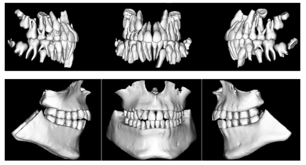
  1. The patient hands your front desk staff a box and an envelope. The box contains a set of plaster study models.  The envelope contains two radiographic films (a panorex and a lateral cephalogram) and a printed sheet with intra and extra oral photographs.
  2. The patient hands your receptionist an envelope. The envelope contains four pieces of paper. Printed on the sheets of paper are the following: intra and extra oral photographs, an image of a panorex, an image of a lateral cephalogram and a picture of a screenshot montage of digital study models.
  3. The patient hands your new patient coordinator a disk. The disk contains several digital files.  Among the files are several JPEGs (digital intra and extra oral photographs), two STL files (3D digital study models with centric occlusion bite registration) and one DICOM file (a large field of view CBCT scan).

What happens next depends on your level of familiarity with digital technologies and which orthodontist’s diagnostic records were presented by the patient.  Another question is whether or not the three different sets of diagnostic records satisfy the AAO’s Clinical Practice Guidelines for Pretreatment Unaltered Diagnostic Records – a full PDF of the Guidelines can be found on the AAO Member Website (Clinical Practice Guidelines).   It is my opinion that in order to best serve our patients, we work to establish guidelines to bridge the analog-digital / 2D – 3D divide and create a standard for maintaining, sharing and transferring both analog and digital diagnostic records.

Where do we start?  Analog diagnostic records (plaster study models / radiographic films) seem fairly straightforward: maintain/provide the originals or a satisfactory copy.  Digital diagnostic records are a different story – 2D or 3D – different manufacturers and different software may format the data files into formats that are incompatible with each other.  For guidance, I turned to the medical community, where digital imaging has been commonplace for decades (Digital Radiography, CT Scans, MRI).  A review of the literature (Exchanging and Sharing Medical Images) provided two important considerations: the imaging files should be the original unaltered files and should be provided in a vendor-neutral format.  We didn’t learn about this in our residency programs!

What’s next?  We already discussed analog diagnostic records.  Here’s my opinion on digital diagnostic records. I feel the original unaltered image files should be handled as follows:

  • Intra / Extra Oral Digital Photographs: Should be provided in a JPEG ( .JPG ) file format.  This format is ubiquitous.  You can view this format on your phone or your office workstations.  All imaging software programs will accept photographs in this format.
  • 2D Digital Radiographs: Should be handled the same as digital photographs: JPEG format.  All imaging software and practice management software should have the capability of exporting and importing digital radiographs in JPEG format.
  • 3D Digital Radiographs (CBCT Scans): The original unaltered DICOM ( .DCM ) files should be provided.  The scan data may be exported as a single .DCM file or as a sequence of multiple (hundreds) .DCM files.  All of the major CBCT scanner manufacturers package their scanners with software that can import / export scan data in this format.  My thoughts on accommodations for a non-3D orthodontist’s use of the DICOM dataset will follow.
  • 3D Digital Study Models: The original unaltered intra-oral scan should be provided in STL ( .STL ) file format.  Typically, the Digital Study Models will be exported from the software as two files that contain the upper and lower models plus a bite registration.  Most major 3D intra-oral scanners contain in their accompanying software the ability to convert and export the raw scan data as an STL (vendor-neutral) file set.  If you own a 3D intraoral scanner that has “closed architecture” and cannot export the scan data in STL format, it is my opinion that you should provide a 3D printed set of study models if asked.  The requirements for diagnostic models in the AAO Clinical Practice Guidelines are clear:  they must be 3 dimensional to suffice.  In my opinion, a printed screenshot of digital study models does not meet these requirements.  There are other considerations in handling digital study models (i.e. open shell/surface scan versus closed shell); however, I feel these are beyond the scope of the current discussion.

Now we must consider how all orthodontists, from non-digital to fully 3D digital, can utilize each other’s diagnostic records.  Let’s look back at our scenarios:

  1. The diagnostic records presented in scenario #1 above do appear to fulfill the requirements in the Clinical Practice Guidelines, except that I would like to see the photographs provided in digital JPEG format as opposed to a printout. If the photographs are indeed “photographs,” I have no advice – except to grab your digital camera, cheek retractors and mirrors.  If you receive these records and feel additional information or imaging is required to provide an accurate diagnosis and treatment plan – that is your (and the patient’s) choice.
  2. In my opinion, the diagnostic records presented in scenario #2 do not fulfill the requirements. Diagnostic models need to be three dimensional – a printout is not.  Regarding the other printed pictures – see my recommendations above.
  3. The disc presented in scenario #3 does, in my opinion, fulfill the requirements. The problem is that a non-3D orthodontist is likely unable to utilize or interpret most of the diagnostic information provided.  How then do we bridge this divide?

In my office where we utilize 3D intra-oral scanning and ultra-low dose CBCT scans, the diagnostic records process is fairly quick and relatively non-invasive.  I’ve yet to have a patient say “I’d rather have an impression” after a 3D intra-oral scan.  I have previously blogged about my opinions on ultra-low dose CBCT scanning (AAO Tech Blog).  I understand that not all orthodontists can utilize, or interpret, the data we produce.  When asked to provide a copy of a patient’s diagnostic records, we produce a disc that contains the following files:

  • Intra and extra oral photographs in JPEG format
  • 3D Diagnostic models – original unaltered – in STL format
  • CBCT Scan – original unaltered – in DICOM ( .DCM ) format: this allows another orthodontist to import the scan data into their own 3D viewing software
  • CBCT Scan – with viewer – provided as a separate proprietary file type: this allows another orthodontist (without their own 3D viewing software) to view the scan data
  • Panoramic view – reformatted as a JPEG file
  • Lateral Cephalogram – reformatted as a JPEG file – with distance ruler for tracing purposes

In my opinion, the disc we produce fulfills the requirements for “Pretreatment Unaltered Diagnostic Records,” and as provided, can be utilized by any orthodontist for diagnostic and treatment planning purposes – with one exception:  the STL files.  If physical study models are requested, the STL files should be printed and provided.  Keep in mind most jurisdictions allow for a nominal copying or duplication fee to be assessed to a patient who requests a copy of their records – the 3D printing charges should fall into this category.

The Committee on Technology will be presenting recommendations to the AAO Board of Trustees in the coming months to aid in amending the Clinical Practice Guidelines in regards to Diagnostic Records, Record Keeping and Transfer of Orthodontic Patients.  My thoughts will be a part of the conversation.  It is my opinion that one day, in the not too distant future, our diagnostic records will be all digital and all in 3D.  As this transition continues, I feel we must take steps to bridge this 2D – 3D / analog-digital divide in a manner that is in the best interest of our patients and within the skillset of every orthodontist.

I ask for your comments and suggestions on this topic.  CTech wants to make sure that all AAO Members are represented in the process as our specialty moves forward with advances in technology.


Sharing Cone-Beam CT Images Online

By Dr. Dan Grauer

When diagnosing and treatment planning interdisciplinary patients, have you ever sent your three-dimensional images to a colleague? Have any of your patients requested a copy of their records for a second opinion? Or maybe, a patient declines a radiograph because another orthodontist has recently taken a CBCT image of the patient? In all of these instances, you will need to communicate with the other office to initiate the transfer of CBCT images. The purpose of this blog is to describe different methods used to share patients’ CBCT records via online means.

Images acquired in your office are requested by a second orthodontist/dentist:

The first question that will need to be answered is whether the other office has the possibility of viewing and analyzing the images in three-dimensions. In a few instances, I have found myself trying to transfer a full three-dimensional file, when the second orthodontist just wanted a cephalogram and a panoramic radiograph. If this is the case, your software will probably allow you to create a synthetic cephalogram and panoramic radiograph that can be emailed through a HIPAA-compliant email account. If the second orthodontist requires a three-dimensional image, two case scenarios are possible:

Case scenario 1: Second orthodontist owns software to read and visualize CBCT images.

In this case, your software is able to export the CBCT Images in DICOM format (Digital Imaging and Communication in Medicine). DICOM files are large, and a file transfer application is needed. Once transferred, these can be imported into the software of the second orthodontist for visualization and analysis.

Case scenario 2: Second orthodontist does not own three-dimensional imaging software.

Under this case scenario, the second orthodontist would need both the CBCT images and a three-dimensional viewer. Three main options are available.

Option 1: If you own a CBCT machine, your software is generally able to create a file that includes both the image data and a basic viewer. The files created are large and can be transferred with a file transfer application.

Option 2: Anatomage offers the possibility of uploading your CBCT images to the cloud, and these can be accessed online through Anatomage’s application, which acts as a visualization tool. At this point the software is in Beta-version and can be accessed at www.anatomagcloud.com. You, as the generating office, will need to upload the images to the AnatomageCloud database and use this application to allow the second office to access the specific patient images. The access is granted with a link embedded in an email. After receiving authorization to access the images, the second office will be able to access the images online without the need of downloading them or installing any software.

Option 3: Dolphin Imaging software offers a complimentary viewer, https://www.dolphinusers.com/dolphin-imaging-viewer/. The receiving doctor can view 3D images by downloading and installing the Dolphin Imaging Viewer software. Files are transferred in DAZ file format. This file format is proprietary to Dolphin Imaging, and the files are created by the originating doctor through Dolphin Imaging 3D Software. This option 3 would work also in Case Scenario 1, when both doctors use Dolphin Imaging 3D software, but it is important to note that only the unprocessed images need to be transferred, such as the DICOM file; the viewer is part of the software downloaded by the receiving office.

Images acquired by other offices:

Images that you receive from other offices should be requested in DICOM format. This will permit you to be able to import these into your 3D software. If you obtain the file in a different format than DICOM (that often includes the viewer), the analysis and measurement possibilities are limited; this is because your 3D software most likely includes all the features that you may need while visualizing and measuring 3D Images. If both offices use Dolphin Imaging 3D Software, a proprietary format DAZ can be used to transfer and share images. The advantage of this approach is that all patient images, including both 3D and 2D images, are shared simultaneously.

In summary, with Cone Beam CT becoming more popular in practices, sharing 3D images with other treating doctors or practices requires some additional steps. The first step is to initiate the conversation with the second office to establish the best system to use to share images. The advantages of 3D images over traditional 2D images are beyond the scope of this blog, but once you become accustomed to a transfer and visualization system, the collaboration between doctors and patient care may improve.

Big Data Revisited

By Anthony M. Puntillo DDS, MSD

In August 2014, I wrote an introductory article for this blog entitled “What is Big Data and How is it Related to the Practice of Orthodontics?” As more orthodontic practices move to the digital collection of orthodontic treatment records (EHRs-photos, models, radiographs, treatment history) and more of our data is being stored in the “cloud”, there is a tremendous opportunity for us as a profession to access that data for the betterment of our patients and advancement of our specialty. Over the last ~3.5 years, however, there has been little visible traction by our researchers and leadership on this front. Meanwhile, there should be no doubt that corporate entities (DSOs and orthodontic vendors) understand the value of our data. Check out the recent cover article for Fortune Magazine (“Tech’s Next Big Wave: Big Data Meets Biology” -3/19/2018). The article notes that “The quest to retrieve, analyze, and leverage (medical) data has become the new gold rush.” If orthodontists are to hope to have any influence on how orthodontic treatment is delivered in the future, management of our patients’ data will be crucial. Technology has sped up every aspect of our lives. We must now start to give this issue the attention it desperately demands. But where should we begin?
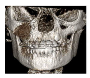
If we are to tackle this challenge, there are many complex questions that will need to be answered. Our patients’ privacy is not the least of these. Even with our busy professional and personal lives, I imagine it has been hard for most to miss the recent public flogging of Mark Zuckerburg and Facebook. Both he and his company were taken to task by Congress and the media when they revealed that the personal data for 87 million of their customers had been inappropriately accessed by an outside research company. As a result of these disclosures, politicians are threatening regulations for their industry – think HIPAA for Silicon Valley. There are more than 2.5 million people who annually seek orthodontic treatment. As we look to find the best ways to utilize our patient’s treatment data to improve their care, we must make certain that it is being done in a way that is respectful of all patients’ privacy. While privacy may be where we start, there are other, even more difficult issues that need to be addressed.

The complexity of the Big Data issue will require the input from the brightest minds both from within and from outside of our profession. To that end, the Great Lakes Association of Orthodontists has put forth a resolution to this year’s American Association of Orthodontists House of Delegates. Resolution 18-18 GLAO (http://hod.live.aaoinfo.org/resolution2/18-18-glao-big-data-task-force-and-records-repository/) requests that our Association President appoint a Big Data task force. I would encourage you to review the resolution and let your representatives (HOD Delegates) know your feelings. While this issue is certain to require a significant investment of time, talent and financial resources, we cannot afford to leave this investment to outside sources. Those who control the data will control the future.

CBCT Imaging for your Practice: Is Now the Time?

By Dr. Kenneth R  Webb

As I walked the exhibit floor at the AAO Annual Session in San Diego this past April, I stopped and talked to several of the company reps displaying the latest generation of CBCT scanners. Competition is good – what struck me is how far this technology has come in such a short period of time. Effective dose, image quality, and versatility are three areas where the advances are most impressive. I have witnessed these advancements first hand in my practice where we have been using CBCT since 2012 and recently upgraded a satellite office from a 2D digital Pan / Ceph to an “Ultra Low Dose” current generation CBCT scanner. I’d like to share some of my thoughts about the 2D to 3D transition.

Effective Dose
I encourage anyone interested in CBCT technology to search for studies authored by Dr. John Ludlow and his team. Dosimetry data and image quality for many CBCT scanners (both older and current generations) have been researched in great detail. Comparative data for 2D dental radiographic imaging (both intra and extra oral) is also available. By replacing our 2D Digital Pan / Ceph with a current generation CBCT scanner we have reduced the effective dose of our diagnostic records imaging by approximately 60%. (Ludlow JB, Walker C. AJO-DO, 2013;144 (6): 802-817) This is accomplished with one, approximately 5 second, ultra-low dose CBCT scan compared to the two longer duration exposures (pan then ceph) required with 2D imaging. Certainly a win-win for our patients.

Versatility
What else can we use the 3D data for? Digital models: used to assess the inter-arch and intra-arch relationship as well as aiding the determination of arch length and arch width requirements and assessing arch symmetry. Modelling labs can produce articulated 3D study models from the CBCT data (DICOM) files.

STL files of the patient’s dentition and occlusion can be uploaded into whatever software you may be using in your office for 3D model storage and viewing. Additionally, these models can be used (with appropriate software) to produce treatment simulations by “moving” individual teeth or the arches as a whole. Imagine completing your diagnostic records with Intra / Extra Oral photos and one 5-second CBCT scan!

Viewing the Diagnostic Data
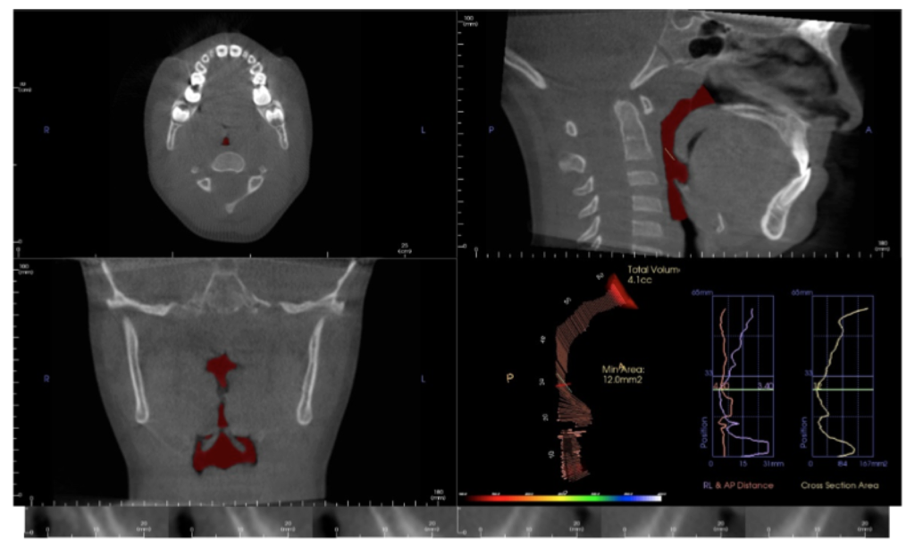
The software that comes with a CBCT scanner can easily produce the customary 2D diagnostic images that we are used to evaluating (panoramic, lateral and A-P ceph). In addition – you see everything! The data can be viewed in sagittal, axial or coronal sections (slices) or as a 3D “volume rendering” which can be rotated and sliced (“clipped”) at will. So why is this additional information gained from CBCT imaging important?

Pathology
You will see pathology in the 3D data that isn’t visible with standard 2D imaging. When pathology is visible in 2D, the 3D data can more accurately ascertain location, extent, and character of the area of concern. This is beneficial to our patients.
Developmental Anomalies
One of my first revelations when we began imaging in 3D was the prevalence and extent of “individual anatomic variation”. But how much is too much? We have made referrals to medical specialists for significant developmental anomalies in the cervical spine, nasal cavity and paranasal sinuses. Patients / parents are appreciative of the thoroughness of our diagnostic process that includes 3D imaging.

Asymmetries
They get their own paragraph! Your patient smiles at you and you notice a vertical asymmetry and occlusal plane “smile” cant. Is it caused by hemimandibular hyperplasia, condylar hyperplasia, a unilateral expansive lesion in the maxilla, a growth response to unilateral progressive condylar resorption (to name a few) or a growth response to a foreign object lodged in the nasal cavity? I’ve seen them all. Similar clinical presentations – different treatment plans.

TMJ
Condylar position in centric occlusion, condylar size and shape, glenoid fossa morphology, condylar osseous morphology, joint space, findings suggestive of degenerative joint disease – both active and stable. It is hard to argue that these are not important considerations in our diagnostic process.

Airway
The value of volumetric and cross-sectional analysis of the naso – pharyngeal airway and its relationship to orthodontic diagnosis and treatment planning has, and continues to be, studied in great detail. Is the growth and development of a patient who presents with narrow arches, an anterior open bite or anterior crossbite, tongue thrust and a strong mouth breathing pattern secondary to adenoid / tonsillar hyperplasia, a deviated nasal septum, environmental allergies or restrictive airway dimensions in general? Should your imaging modality include an assessment of these areas?

And there is more…
Orthognathic surgical treatment planning, TAD placement guidance, precise localization of ectopic and supernumary teeth, and assessment of treatment progress – including evaluation of root torque. All are possible or enhanced with 3D imaging. And by managing scan parameters (field of view, scan time and voxel size) at an effective dose equal to or less than 2D imaging modalities.

So, if you haven’t brought this technology into your practice – is now the time?

At the 2017 AAO Winter Conference in Ft. Lauderdale, Mr. Chris Bentson reported on a survey of recent (2016) U.S. Orthodontic Residency graduates: 88% responded that they had used CBCT imaging for diagnosis and treatment planning during their residencies including 21% that used it on all patients.

For the 2013 graduates, the responses were 76% and 9% for the same questions.

Based on these statistics, are we that far away from 3D imaging being included in the “standard of care” discussion? Embracing new technology is not easy but the value added for the practitioner and our patients is significant. The orthodontic educators who expertly taught my generation faced a similar situation during their careers with a then relatively new technology: panoramic radiographic imaging. My generation faced the challenges of transitioning from analog imaging (film) to digital.

Change is not easy. The transition from 2D to 3D imaging in your practice will require a financial and educational commitment. The whole staff will be a part of this paradigm shift. A positive – 3D imaging will be a differentiator for your practice. If you are seeking an associate, partner or complete transition, 3D imaging will make your practice more attractive to the next generation of orthodontists.

3D imaging will benefit you and your patients. Is now the time?

Is it Time to Say Goodbye to our DSLR Cameras?

By Dr. Anthony M. Puntillo, DDS, MSD

When I first graduated from my orthodontic residency, now more than 23 years ago, a standard set of initial records included plaster models, facial and intraoral photographs taken with film, developed, then trimmed and placed into mounts and panoramic and lateral cephalometric radiographs taken with film and developed in darkrooms. The digitization of our society has made the process of gathering and storing this important diagnostic information much more efficient for most orthodontists. In fact, more than four years ago (November 2012) I wrote a Tech Blog article on digital retainers and the impressionless orthodontic practice. Since then the use of intraoral scanners and 3D printing in our profession has grown exponentially. It is now not hard to imagine a day in the near future when impressions will disappear completely from the practice of dentistry.  As I near the end of my 8 year term on the CTECH committee, I can’t help but wonder what is next.   Where else can we use technology to eliminate inefficient processes from our practices?

The most obvious next step for me is the elimination of intraoral photographs. All of those intraoral scanners, now used by most orthodontists, take multiple photographs of our patients’ teeth to create the 3D digital images. Several of these scanners can capture images in true, or close to true color. It can’t be long before we come to the realization that digital images taken with a good intraoral scanner are a better alternative to the standard set of 5-7 intraoral 2D photos we have been taking for decades. The 3D digital image is not only a better diagnostic record of the patient’s current dental state, it also is more versatile in that it can also be used to create and fabricate appliances (i.e. clear aligners, indirect bonding setups, retainers, etc.). If a good intraoral scan can consistently be completed in less than 10 minutes, aren’t we wasting our time and that of our patients’ taking 2D photos. I concede that we are all very accustomed to diagnosing our patients with these 2D photographic images. However, it was not that long ago when most thought that multiple radiographic exposures were necessary on the majority of our patients.   Now most of our patients are diagnosed with a single, quick radiograph taken on a CBCT machine and from that single exposure we derive a much higher level of diagnostic information.

To be honest, I am not yet ready to mothball our cameras. For starters, I still think that facial 2D photos are necessary. I know that there are 3D cameras available that will someday eliminate the need for our extraoral series of facial photos. However, for whatever reason (I believe primarily cost) these have not yet caught on. So for now we will still be taking a series of three 2D digital photos of our patients’ faces. Additionally, 2D intraoral pictures still play a significant role in our new patient consultations. We have not yet found the best way to display and share the captured 3D dental images (STL files) to educate our patients. I anticipate that this last hurdle will be overcome in 2017 and when that happens our DSLRs are going to see much less action and our IOSs are going to play an even larger role in our new patient process.

3D Digital Indirect Bonding…Why You Should Consider It for Your Practice

By Dr. Ed Lin

DrLin-Headshot-2Indirect bonding was first introduced to orthodontics over 20 years ago and has become an integral part of many orthodontic practices and orthodontic labs worldwide. The four reasons/advantages for its inception are: 1) Indirect bonding has been widely viewed as giving the orthodontist the ability to achieve more accurate bracket placement on a static model and not having to deal with the clinical challenges with direct bonding on a patient, 2) The doctor can perform the final check for bracket placement at his/her own leisure and not under a set clinical schedule, 3) To improve clinical efficiency with decreased doctor chair time at the full bonding appointment, and 4) Finally, for improved patient comfort due to decreased time which the patient is in cheek retractors.

Having utilized indirect bonding in our practices for over 17 years now, these four reasons/advantages are without a doubt why all four doctors in our practices utilize indirect bonding for all of our patients. However, there are some disadvantages that are also present with conventional indirect bonding such as: 1) Distortion of the impressions leading to inaccurate models/trays, 2) Distortion of the pour up of the impressions resulting in inaccurate models/trays, 3) Short clinical crowns are not ideal for conventional indirect bonding and need to be direct bonded especially in the mandibular arch, 4) Brackets can be bumped prior to curing on the model leading to incorrect bracket placement, and 5) Seating of the indirect bonding tray is technique sensitive and can lead to over seating or under seating of the trays resulting incorrect bracket placement.

With the advancements of technologies in dentistry with intraoral scanning, 3D digital virtual bracket placement software, and 3D printing, these technologies have now given us the capabilities in orthodontics to improve upon conventional indirect bonding with 3D digital indirect bonding. Currently, there are 4 companies that I am aware of in the US market that offer 3D digital indirect bonding: OrthoSelect’s Digital Indirect Bonding System (DIBS), Arcad Digital Indirect Bonding System, Great Lakes Orthodontics’ Exceed, and SureSmile’s Elemetrix 3D Printed Indirect Bonding System. All four of these companies have proprietary cloud based software for management of patient case submissions and all of them accept intraoral scans.

For Arcad and Great Lake Orthodontics, upon final approval of the virtual bracket positions in their proprietary software, they will 3D print the models with specific indexes for very precise and accurate bracket placement on the 3D printed models. Once the brackets are placed manually on the 3D printed indexed models, their labs will then create an indirect bonding tray through conventional lab techniques and are then ready to be utilized for indirect bonding for the patient. The advantages with both systems are that they are dealing with digital data with an intraoral scan so there are no issues with a poor impression. Also, with 3D printed models, there are no issues with a poor model with air bubbles or blebs on the model. As a result, the conventional indirect bonding trays are more accurate and precise.

For OrthoSelect’s DIBS and SureSmile’s Elemetrix indirect bonding systems, upon final approval of the virtual bracket positions in their proprietary software, both of these companies will 3D print the indirect bonding trays. The brackets are then manually placed into the 3D printed indirect bonding trays for both systems and are then ready to be placed on the patient. OrthoSelect’s DIBS and SureSmile’s Elemetrix systems have the same 2 advantages as Great Lakes and Arcad’s systems, as they are only dealing with digital data with an intraoral scan, so there are no issues with poor impressions or poor model pour ups. However, since only the indirect bonding trays are 3D printed, both OrthoSelect’s and SureSmile’s systems have 2 advantages over Great Lakes and Arcad – 1) There is a significant time savings on the lab side of things since there are no 3D printed models, only the indirect bonding trays are 3D printed and 2) There is no human error brought into production of the indirect bonding trays since they are 3D printed.

Our practices are now making the transition from conventional indirect bonding to 3D digital indirect bonding with SureSmile’s Elemetrix 3D printed indirect bonding tray system so I will give a brief overview of how we utilize this in our practices. We have a 3 Shape Trios 3 intraoral scanner at each practice location. With the “Insane Mode” of the Trios, we can intraoral scan 2 arches with a digital bite registration in a total of 1 minute. We then upload the intraoral scan through the cloud to SureSmile. SureSmile will then segment, clean up, and create the 3D digital model and we will receive the digital model back on our end in 1-2 business days. We then assign the virtual brackets (.018 American Empower stainless steel or clear) and virtually position them on the digital models. Upon final approval of the virtual bracket placements, we then order our SureSmile 3D printed indirect bonding trays. The indirect bonding trays are then 3D printed and then shipped to us. We currently schedule 15 days between the intraoral scan for indirect bonding and the full bonding appointment to allow time for setup and approval, 3D printing of the trays, and for shipment to us. In the future, I hope that we will have the capabilities to 3D print our own indirect bonding trays in house, which will give us the capabilities to have quicker turnaround time with the indirect bonding trays and we will not have to deal with any shipping issues. Because SureSmile’s 3D printed indirect bonding trays are rigid, there is also an added benefit with their indirect bonding trays as we do not have to worry about over seating or under seating of their trays as you do with conventional indirect bonding trays, which are softer, as a result of the silicone and polyvinyl siloxane materials which are used to make them. The total cost for a full indirect bonding case with SureSmile’s Elemetrix Indirect Bonding System is $105 which is very affordable and attractive to me especially considering the added benefits which I have stated above.

My personal opinion is that any of the 4 digital indirect bonding systems, which I have stated above, are a step above and are an improvement upon any conventional indirect bonding systems currently available. If you are currently utilizing conventional indirect bonding or are considering transitioning to indirect bonding in your practice, I would strongly encourage you to evaluate the digital indirect bonding systems which are currently available today. As with all other aspects of our lives, technology is changing the way we all live and is helping to improve how we do things in everyday life as well as with our practices.

eOrthodontics

By Dr. Anthony M. Puntillo, DDS, MSD

Dr.-Puntillo-PictureIt was not that long ago when we all relied upon our friendly postmen and postwomen for the delivery of our letters. Today the United States Postal service is scaling back mail operations in favor of package delivery, and the majority of our written communication is transmitted electronically. Is the delivery of healthcare, and particularly orthodontic care, headed for a similar fate? A Computerworld article, cited research by Deloitte, which projected 75 million of 600 million appointments in 2014 with general practitioners would involve electronic or eVisits¹. “Electronic visits or telemedicine are comprised of electronic document exchanges, telephone consultations, email or texting, and videoconferencing between physicians and patients. The vast majority of eVisits, according to Deloitte, are likely to focus on capturing patient information through electronic forms, questionnaires and photos.” In the state of Texas, new legislation has opened the door for physicians to be compensated for remotely providing care to children through a video connection to the school nurses’ office². Market forces including an expansion of access to care, increased efficiency, and financial incentives are driving all of these changes. Just as eMail though has not completely eliminated the need for our postal service, eVisits are not likely to eliminate the need for all direct patient to physician interaction. However, there can be no denying that technology is changing the manner in which healthcare is delivered and our specialty will not be immune.
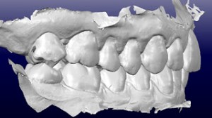
It may be hard to conceive how we will remotely align teeth. However, the 1999 introduction of digitally created tooth moving clear aligners (Align Technology, Inc.) opened the door to the Orthodontic digital age and fortunately or unfortunately that door cannot be closed. Recently, while attending an orthodontic meeting overseas, I noticed a new company that seemed to be garnering a great deal of attention, Dental Monitoring (dental-monitoring.com). This company claims to be the “first monitoring solution in orthodontic care.” Patients capture their tooth movements with their smartphones and orthodontists can remotely monitor and direct treatment via an app. Whether or not this company is the first is irrelevant. It will certainly not be the last. The same efficiencies and cost advantages that eVisits offer physicians in the general practice of medicine also apply to orthodontic practices. To be clear, I am not advocating that remote care is better or even desirable. The purpose of this blog is to raise our members’ awareness and open discussions regarding technological changes in our profession. In the last 20 years, we have incorporated electronic patient records, CBCTs, intraoral scanners, and even 3D printing into our practices. As we all adapt to the expectations of the Millennial Generation what will our practices look like 20 years from now?

1. Almost one in six doctor visits will be virtual this year. Computerworld: Mearian, L. August 8, 2014.
2. Law could bring remote doctor visits to schools. The Texas Tribune: Rocha, A et. al., August 23, 2015.

Smartphone-based orthodontic monitoring: the big brother in our patient’s mouth

by Domenico Dalessandri, DDS, MS, PhD

Screen Shot 2016-03-11 at 11.06.09 AMNowadays almost all of us have a smartphone and we use it hundreds of times during the day checking incoming e-mails, surfing through the web and sharing our thoughts and pictures online within our social communities. This is the “permanently online” era and our patients ask for immediate information supply and assistance request satisfaction.

Since sometime orthodontists have used text messaging apps like WeChat or WhatsApp to communicate with their patients, to remind them the next visit, to solve an emergency or replying to a late doubt, or even checking the oral hygiene status or the cooperation level with removable appliances. There is a growing evidence in the literature showing the efficacy of these “App based” monitoring protocols in ameliorating oral hygiene and reducing treatment duration, bracket bond failure, and failed or late attendance [1-2].

Commercial interest regarding patient monitoring has also increased progressively and nowadays there are few apps especially designed for this purpose. In all these systems the orthodontist has his own dashboard on reserved area in the company website that can be used to invite patients to download the app in their smartphones and to visualize their smile pictures.

SmileTrackerTM (TP Orthodontics) also allows visualizing a time-lapse video, based on daily pictures taken by the patient during the monitoring, of the treatment progress. Furthermore this app has a rewards-based system in order to keep patients engaged throughout the monitoring and it also allows them to share their progresses through their own social profiles.

Dental Monitoring® allows patients to take smile pictures every 2 weeks in case of fixed orthodontic treatment or even every week for invisible aligners treatments. Doctors can also ask for a supplementary exam whenever they want, sending through their personal homepage a specific invitation that is forwarded by e-mail to the patient. This system requires doctors to send patient dental impressions before monitoring begins, because it utilizes a specific algorithm that allows calculating teeth movements based on pictures 3D matching and superimposition on the initial virtual models. For this reason patients are required to take 13 pictures for each exam from different angulations focusing on both single arches and on their position in occlusion. Three more pictures are required for patients treated with invisible aligners, in order to check each single aligner fit. This system, apart from calculating through the algorithm tooth by tooth displacement as mesial/distal, intrusion/extrusion, retraction/advancement, rotation, inclination and angulation variations, provides pictures observation by an orthodontist that looks at oral hygiene level, aligners fitting, possible presence of teeth abrasion, decays or brackets failure, sending a warning to the treating orthodontist in case of any unexpected event.

Could these systems become important tools allowing us to improve our treatments efficiency and quality? Is it credible that “virtual” appointments could in some occasions substitute the traditional “physical” appointments? Will our patients be available to be continuously controlled by this orthodontic “big brother”?

It is hard to foresee the future. The possibility for patients, especially if they live far from the orthodontic office, to save time and money by reducing the number of visits having the same, or even a better, accuracy in treatment progress control, can be universally recognized as positive factors fostering the diffusion of these systems.

Are we ready for this further step forward along the digitalization path of our beloved orthodontic specialty?

1: Zotti F, Dalessandri D, Salgarello S, Piancino M, Bonetti S, Visconti L, Paganelli C. Usefulness of an app in improving oral hygiene compliance in adolescent orthodontic patients. Angle Orthod. 2016;86:101-7.

2: Li X, Xu ZR, Tang N, Ye C, Zhu XL, Zhou T, Zhao ZH. Effect of intervention using a messaging app on compliance and duration of treatment in orthodontic patients. Clin Oral Investig. 2015 Dec 2. [Epub ahead of print]

Domenico Dalessandri qualified in Dentistry from the University of Brescia, Italy, where he received specialty training in Orthodontics. He obtained his PhD from the University of Torino discussing a thesis entitled “Cone Beam Computed Tomography: accuracy and reliability”. He received a research fellowship in “CBCT applications in Orthodontics” by the University of Trieste. Currently he is Adjunct Assistant Professor at the University of Brescia.

He has additional clinical research interests in: indirect bonding; self-ligating braces; use of mini implants and miniscrews; lingual orthodontics; Invisalign; cleft lip and palate; impacted teeth; 3D technology and virtual treatment planning; CAD-CAM; corticotomy and piezocision.

3D Printing and Orthodontics

By Dr. Christian Groth

As we move towards the 2015 AAO Annual Session in San Francisco many of us will be making check lists for items to investigate at the exhibition hall and lectures. Anybody who has attended recent meetings has seen that intraoral scanners are a hot topic. Every year new products are being released, or updates to current systems are offered. Intraoral scanning has opened the door for additional technologies within (and outside of) the orthodontic office. Dr. John White wrote a very informative blog post in February talking about the use of intraoral scanning for same day consultations (click here to read it). As more people are offering clear aligner therapy (including general dentists and the mail order aligner system that we all know about) it is time that we differentiate ourselves as orthodontic specialists. One way in which we can do this is to incorporate 3D printing into our daily practices.

3D printing, also known as additive manufacturing, is a process by which a physical object is created from a digital file (check out a video of 3D printed models here). There are several different types of 3D printers available that range in price from a few hundred dollars to almost one hundred thousand dollars. They all have one thing in common: they build models layer-by-layer with a build platform that moves vertically. The smaller the layer thickness the better looking the model will be. The four most popular types of printers are: Fused Deposition Modeling (FDM), Stereolithography (SLA), Digital Light Projector (DLP), and Polyjet Photopolymer (PP). Without getting too technical here is how each basically works. FDM involved heating up a thin strand of plastic resin that comes off of a spool and is deposited in layers as thin as 100 microns. SLA and DLP technologies are similar in that they utilize a vat of liquid, photosensitive resin. When the light hits the resin it is cured and platform moves to enable the next layer to be cured. The different between SLA and DLP is that SLA uses a single laser point to draw an image whereas DLP uses a projected image to cure a whole layer simultaneously, which allows the printing process to move faster (think of this as the difference between drawing a picture and stamping a picture). PP printers are probably the most popular in dentistry and use inkjet technology (yes, just like your desktop printer). Liquid resin is jetted out of nozzles in an extremely accurate fashion and cured by a UV. Layer thickness of SLA, DLP, and PP printers can be as low as 16 microns (for your reference the average piece of paper is 100 microns thick).

While the technologies differ between printers what truly sets them apart is the quality of the parts. Cheap printers are made from cheap parts that can degrade over time and result in inaccurate models. It is truly a case of you get what you pay for. So if you are in the market for a 3D printer to be used in your practice, buy the best one that you can afford.

3D printed models can be used for anything in the orthodontic office.   The most practical use is for retention and relapse treatment. A major downside to stone models is that they are often destroyed during the retainer fabrication process. 3D printed models do not get ruined and can be used as many times as necessary for retainer fabrication. Imagine being able to print a model, make a clear retainer, and mail it off to a patient who is away at college. What a service you have just provided to your patient and they never stepped foot in your office! Pairing 3D printing with one of several software programs available allows us to create sequential setups/models for minor tooth movement. By controlling the process we control the overhead and thus have the ability to pass these savings on to the patient. Another great service that we can offer our patients if they have a lapse in retainer wear.

Whatever your practice is like there is a place for 3D printed models in it. While you will pay a little bit more for the physical model, the longevity, versatility, patient excitement, and ability to virtually eliminate alginate impressions from your practice will pay off in the end!